Sars-Cov-2 PCR

Background

During the peak of the COVID-19 pandemic in Kazakhstan (June 2020), the media reported multiple cases of SARS-CoV-2 PCR-negative pneumonia with increased mortality. Our objective was to study the epidemiological characteristics of hospitalized patients with positive and negative PCR with analysis of hospital and post-hospital mortality. We also compare the characteristics of respiratory diseases between 2019 and 2020.

Methods

The study population consists of 17,691 (March-July-2020) and 4,600 (March-July-2019) hospitalized patients with respiratory diseases (including COVID-19). Incidence rate, case fatality rate, and survival analysis for overall mortality (in-hospital and post-hospital) were evaluated.

  • Study population and data sources

The study population consisted of all hospitalized patients with respiratory illnesses (including COVID-19) according to the International Statistical Classification of Diseases and Related Health Problems (ICD-10) from March to July 2019 and from March to July 2020 in Turkestan oblast, Kazakhstan. The following ICD-10 codes were included in the study: J00-J06 (acute upper respiratory tract infections), J09-J18 (influenza and pneumonia), J20-J22 (other acute lower respiratory tract infections), J40- J47 (chronic diseases of the lower respiratory tract), J96-J99 (other diseases of the respiratory system), B34 (viral infection of unspecified site), Z20 (contact and “suspected” exposure to communicable diseases), U07.1 (COVID-19 specified virus) and U07 .2 (COVID-19 unspecified virus).

The raw data was retrieved from the Single National Electronic Health System (UNEHS) linked with the records to the “Electronic Registry of Internal Patients” that included data on dates of admission and discharge, ICD-10 codes, dates and results of PCR tests, discharge results and some demographic data. Global mortality statistics (in-hospital and post-hospital death) were obtained independently from the “Adjunct Population Registry” and were linked to hospitalized patients through the Population Registry Number (RPN-ID); each date of death followed by the date of hospital discharge is considered post-hospital mortality. The population census of the Turkestan oblast, including all cities and rural areas (2,016,100 people), was obtained from the State Statistics Committee.

  • SARS-CoV-2 infection detection method

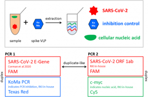
Confirmation of SARS-CoV-2 infection was performed by real-time quantitative PCR on nasopharyngeal swabs with the BGI kit (Beijing Genomics Institute, Shenzhen, China) in defined special regional laboratory settings.

  • Assessment results

Incidence, mortality and lethality rates were evaluated. Incidence and mortality rates were calculated for each year using the number of newly diagnosed patients and deaths, and the size of the population. The case fatality rate was calculated by dividing the number of deaths by the number of newly diagnosed cases. The incidence was compared by year of admission. All-cause mortality was divided into in-hospital and post-hospital mortality, which was used to identify associated risk factors among admissions in 2020.
The start of follow-up was the date of hospital admission, and patients were followed until death or the end of the follow-up period (August 30, 2020). Two outcome variables were of interest for survival analysis: in-hospital mortality (time from hospital admission to hospital discharge) and overall (in-hospital and post-hospital combined) mortality (time from hospital admission to death at any time up to 30 days). August 2020). ). Censoring for in-hospital mortality survival analysis was taken on the date of hospital discharge, and for pooled mortality, it was August 30, 2020.

  • Statistic analysis

For each diagnostic group, absolute numbers of hospitalizations and deaths, incidence and mortality rates per 100,000, case fatality rates per year were reported. Absolute and relative frequencies were reported for categorical variables. Means and standard deviations were used to describe continuous variables, while biased continuous variables were characterized by medians and interquartile ranges (IQRs). Parametric bivariate analysis (Pearson’s Chi-squared, two-sample t-test, ANOVA) was used to assess associations of demographic and disease-related characteristics with outcome variables.

Kaplan-Meier survival curves were plotted for the results of the PCR test. Cox proportional hazards models were fitted with epidemiologically and statistically significant covariates using backwards stepwise selection. The proportional hazards assumption for different groups was tested using log plots. We performed a sensitivity analysis to assess the robustness of our main findings.

We examined the association between overall mortality (in-hospital and post-hospital) and sociodemographic parameters in a subgroup of patients admitted to only provisional and infectious disease hospitals (excluding patients who were in quarantine). The significance level of 5% (α < 0.05) was taken. All statistical analyzes were performed using STATA 16.0 statistical software. The study was approved by the Institutional Review Ethics Committee (NU-IREC 203/29112019) with exemption from informed consent.

Results

Respiratory disease incidence and mortality rates were 4 and 11 times higher in 2020 compared to 2019 (877.5 vs. 228.2 and 11.2 vs. 1.2 per 100,000, respectively). PCR-positive cases (compared to PCR-negative) had a two-fold increased risk of overall mortality. We observed a 24% higher risk of death in men than in women and in older patients than in younger ones. Patients residing in rural areas had a 66% higher risk of death compared to city residents, and being treated in a makeshift hospital was associated with 1.9 times higher mortality compared to those treated in hospitals of infectious diseases.

Conclusion

This is the first study from the Central Asia and Eurasia regions, assessing mortality from SARS-CoV-2 PCR positive and PCR negative respiratory system diseases during the peak of the COVID-19 pandemic. We describe a higher mortality rate for PCR-positive cases compared to PCR-negative cases, for men compared to women, for older patients compared to younger patients, and for patients living in rural areas. rural compared to city residents.

SARS-CoV-2 Spike Variant

Summary

Although most mutations in the severe acute respiratory syndrome coronavirus 2 (SARS-CoV-2) genome are expected to be deleterious and rapidly cleared or relatively neutral, a small proportion will affect functional properties and may alter infectivity. , the severity of disease, or interactions with the host. immunity. The appearance of SARS-CoV-2 in late 2019 was followed by a period of relative evolutionary stasis that lasted approximately 11 months.

However, since the end of 2020, the evolution of SARS-CoV-2 has been characterized by the appearance of sets of mutations, in the context of “variants of concern”, that affect the characteristics of the virus, including transmissibility and antigenicity, probably in response to the changing immune system profile of the human population. There is emerging evidence of reduced neutralization of some SARS-CoV-2 variants by post-vaccination serum; however, a greater understanding of the correlates of protection is required to assess how this may affect vaccine efficacy.

However, manufacturers are preparing platforms for a possible update of vaccine sequences, and it is critical that monitoring of genetic and antigenic changes in the global virus population be carried out alongside experiments to elucidate the phenotypic impacts of vaccines. mutations. In this review, we summarize the literature on mutations of the spike variant protein of SARS-CoV-2, the primary antigen, focusing on their impacts on antigenicity and contextualizing them in protein structure, and discussing them in the context of frequencies of mutation observed in the world sequence data sets.

SARS-CoV-2 spike variants

Sites of variation in the SARS-CoV-2 spike protein. Amino acids in bright red have variations in many individuals, pink amino acids vary in fewer individuals, and white amino acids show very few variants. Viruses, in their nonsensical way, are masters of evolution. Two aspects of viral biology make them particularly successful. First, huge populations of viruses are generated as they infect cells and replicate. For example, during the peak of SARS-CoV-2 infection, there may be between 1 and 100 billion viruses in an infected person.

Second, their molecular machinery for replication is often sloppy, introducing occasional errors into the progeny. This is the perfect combination for rapid evolution. During an infection, many variants of the virus can be produced in these populations. Most sequence variations will harm the virus or be neutral with little change for better or worse, but the occasional variant will improve some aspect of the viral life cycle. These rare advantageous variants have emerged several times in SARS-CoV-2 and have caused new waves of infection in the current COVID-19 pandemic.

Variation assessment

Scientists around the world have studied the evolution of SARS-CoV-2 to understand its capabilities and help plan for the future. The illustration shown here maps the main sites of variation of the spike protein, based on more than 3 million samples that have been sequenced and deposited in the GISAID database. The structure is based on PDB ID 7kj2, but the coordinates were taken from SWISS-MODEL since the original PDB entry does not have atomic coordinates for several flex loops. Also, glycosylation is not shown in this illustration, to make protein variation easier to see, so you should imagine the protein covered with multiple carbohydrate chains.

Functional improvements

As you can see, the variation sites are scattered throughout the three-dimensional structure. Scientists are still working out the functions of each of these changes, but some of the more common sites of variation are becoming clearer. The most common mutation (currently at least) is at position 614.

It is believed to control the stability of the top of the spike. Another common mutation, 681, is found in a flexible loop that is clipped by the cellular protease furin, breaking the chain into two pieces. The upper part (S1) recognizes the host cell and the lower part (S2) directs fusion and cell entry. Researchers have found that this cleavage makes the virus more infectious with cells in the respiratory tract.

Variant structures

During the COVID-19 pandemic, SARS-CoV-2 has spread throughout the world and variants have emerged by chance in different countries and spread rapidly from there. Recent variant structures (PDB IDs 7lwv, 7lyo, 7v7q, 7v7e, 7t9k). They all have multiple changes, including sites where an amino acid has mutated (shown in red) and sites where amino acids have been removed from the chain.

They all include the two common changes mentioned above, along with other changes scattered throughout the structure. These can benefit the virus in many ways: mutations in the receptor-binding domain and C-terminal domains can improve recognition and attachment to cells, changes in the N-terminal domain can help evade the immune system and mutations in the S2 region can enhance the process of fusion and cell entry.

Peak variation at position 614

Mutation from aspartate to glycine at position 614 (shown in red) removes an interaction with threonine 859 (turquoise) in a neighbouring subunit in the trimeric peak. This is thought to loosen the structure, facilitating the transition to the active conformation with extended receptor-binding domains. To compare the native structure with aspartate at position 614 (PDB ID 6vyb) and the variant delta-mutated structure with glycine (PDB ID 7v7q).

Nasopharyngeal Carcinoma

Overview

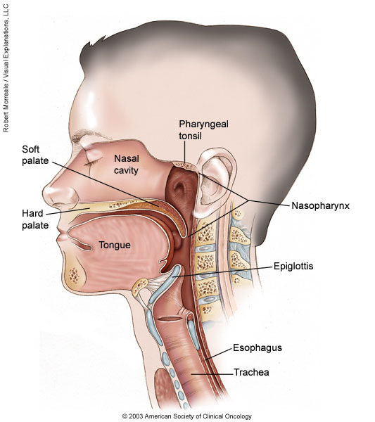
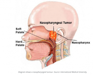
Nasopharyngeal carcinoma is a cancer that occurs in the nasopharynx, which is located behind the nose and above the back of the throat. Nasopharyngeal carcinoma is rare in the United States. It occurs much more frequently in other parts of the world, specifically in Southeast Asia.

Nasopharyngeal carcinoma is difficult to detect early. This is probably because the nasopharynx is not easy to examine and the symptoms of nasopharyngeal carcinoma are similar to other more common conditions. Treatment for nasopharyngeal carcinoma usually involves radiation therapy, chemotherapy, or a combination of the two. You can work with your doctor to determine the exact approach for your particular situation.

Symptoms

In its early stages, nasopharyngeal carcinoma may not cause any symptoms. Possible notable symptoms of nasopharyngeal carcinoma include:

  • A lump in the neck caused by a swollen lymph node
  • blood in your saliva
  • Bloody discharge from the nose
  • Nasal congestion or ringing in the ears
  • Hearing loss
  • Frequent ear infections
  • Throat pain
  • Headaches

When to see a doctor

Early symptoms of nasopharyngeal carcinoma may not always prompt you to see your doctor. However, if you notice unusual and persistent changes in your body that don’t seem right to you, such as unusual nasal congestion, see your doctor.

Causes

Cancer begins when one or more gene mutations cause normal cells to grow out of control, invade surrounding structures, and eventually spread (metastasize) to other parts of the body. In nasopharyngeal carcinomas, this process begins in the squamous cells that line the surface of the nasopharynx.

It is not known exactly what causes the genetic mutations that lead to nasopharyngeal carcinoma, although factors, such as the Epstein-Barr virus, have been identified that increase the risk of this cancer. However, it is not clear why some people with all risk factors never develop cancer, while others with no apparent risk factors do.

Risk factor’s

Researchers have identified some factors that seem to increase the risk of developing nasopharyngeal carcinoma, including:

  • Sex. Nasopharyngeal carcinoma is more common in men than in women.
  • Race. This type of cancer most commonly affects people in parts of China, Southeast Asia, and North Africa. In the United States, Asian immigrants have a higher risk of this type of cancer than Asians born in the United States. Alaskan Eskimos are also at increased risk of nasopharyngeal cancer.
  • Years. Nasopharyngeal cancer can occur at any age, but it is most often diagnosed in adults between the ages of 30 and 50.
  • Salt-cured foods. Chemicals released in the steam when cooking salt-cured foods, such as canned fish and vegetables, can enter the nasal cavity, increasing the risk of nasopharyngeal carcinoma. Being exposed to these chemicals at a young age can further increase the risk.
  • Epstein Barr virus. This common virus usually produces mild signs and symptoms, like those of a cold. It can sometimes cause infectious mononucleosis. The Epstein-Barr virus is also linked to several rare cancers, including nasopharyngeal carcinoma.
  • Family history. Having a relative with nasopharyngeal carcinoma increases the risk of developing the disease.
  • Alcohol and tobacco. Excessive alcohol consumption and tobacco use can increase the risk of developing nasopharyngeal carcinoma.

Complications

Complications of nasopharyngeal carcinoma can include:

  • Cancer that grows to invade nearby structures. Advanced nasopharyngeal carcinoma can cause complications if it grows large enough to invade nearby structures, such as the throat, bones, and brain.
  • Cancer has spread to other areas of the body. Nasopharyngeal carcinoma often spreads (metastasizes) beyond the nasopharynx.

Most people with nasopharyngeal carcinoma have regional metastases. This means that cancer cells from the original tumour have migrated to nearby areas, such as the lymph nodes in the neck. Cancer cells that spread to other areas of the body (distant metastases) most often travel to the bones, lungs, and liver.

Prevention

There is no sure way to prevent nasopharyngeal carcinoma. However, if you are concerned about your risk of nasopharyngeal carcinoma, you may want to consider avoiding habits that have been associated with the disease. For example, you can choose to reduce the amount of salt-cured foods you eat or avoid these foods altogether.

Tests to detect nasopharyngeal carcinoma

In the United States and other areas where the disease is rare, routine screening for nasopharyngeal carcinoma is not done. But in areas of the world where nasopharyngeal carcinoma is much more common—for example, in some areas of China—doctors may offer screening to people thought to be at high risk for the disease. Screening may include blood tests for the Epstein-Barr virus.